
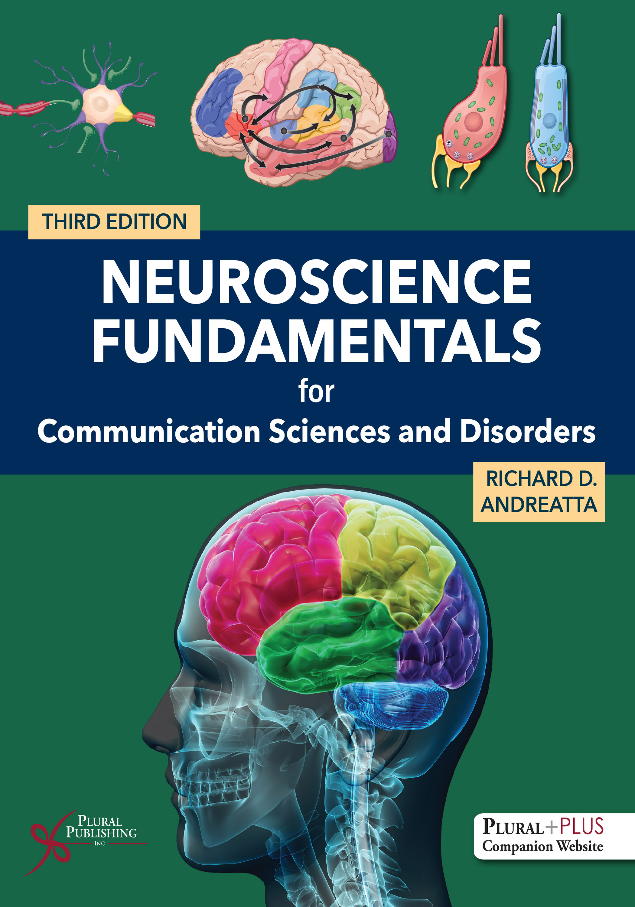
Neuroscience Fundamentals for Communication Sciences and Disorders
Third Edition
Richard D. Andreatta
Details: 793 pages, Full Color, Hardcover, 8.5" x 11"
ISBN13: 978-1-63550-782-9
© 2028 | Available
For Instructors
Purchase
Neuroscience Fundamentals for Communication Sciences and Disorders, Third Edition continues to set the standard for teaching the neural bases of communication with a uniquely engaging, clinically relevant, and student-friendly approach. Designed for undergraduate neuroanatomy and graduate neuroscience courses in CSD programs, this comprehensive text also serves as a trusted reference for practicing speech-language pathologists and audiologists in medical and rehabilitation settings.
Written in a conversational style that incorporates humor, relatable analogies, and clear explanations, the text demystifies complex neuroanatomy and neurophysiology while maintaining scientific rigor. More than 350 beautifully rendered, full-color illustrations bring key concepts to life, supporting visual learning and helping readers make meaningful connections between brain structures, neural processes, and communication outcomes.
The Third Edition builds on its strong foundation with a more intuitive organization, expanded clinical content, and enhanced learning tools that foster critical thinking and clinical application. By emphasizing the integration of brain and behavior, this text equips students and practitioners with the knowledge and confidence to interpret neurological information and apply it to assessment and intervention in real-world settings.
Whether used in the classroom or in clinical practice, Neuroscience Fundamentals for Communication Sciences and Disorders provides a clear, cohesive, and clinically meaningful understanding of the human nervous system as it relates to speech, language, and hearing.
New to the Third Edition
- Addition of 45 detailed clinical case studies, strategically integrated throughout the text to strengthen structure–function–disorder connections and teach clinical reasoning
- Over 80 summary tables, including nearly 40 new ones, to support efficient review and consolidation of key concepts
- Extensive reorganization of content into four new thematic sections for better progression and integration across topics
- Division of longer neuroanatomy chapters into shorter, more digestible, and logically sequenced content
- A new chapter on neural mechanisms of speech and language, with streamlined theoretical discussions and expanded clinical coverage of neurologically based communication disorders
- Separate speech, language, and hearing chapters from the previous edition are now embedded within neuroanatomical and systems chapters for better structure–function–behavior integration
- Reorganization of auditory system chapters for clearer peripheral-to-central progression and improved sequencing
- Restructuring of motor systems content, including a new unified chapter linking muscle contraction with direct motor control and a separate chapter dedicated to indirect motor systems
- Consolidation of neuroplasticity and sensorimotor learning content into a final streamlined capstone chapter, emphasizing behavioral adaptability and sensorimotor learning
PluralPlus Online Ancillaries
For instructors: PowerPoint Slides, Test Bank, Image Bank (with labeled and unlabeled figures), Syllabus with different reading schedules to accommodate varying semester lengths
For students: Chapter Study Guides, Study Question Worksheets, Cranial Nerve Testing Guide, eFlashcards, Unlabeled Anatomy Illustrations, Brain Model Demonstration Videos, Mind Maps, Simplified Summaries on Technical Content.
The Third Edition now includes Brain Model Demonstration videos! View a sample featuring the cortex:
Contents
Preface
About the Author
About the Illustrator: Maury Aaseng
Contributors
Reviewers
Acknowledgments
Section 1. Foundations: Neuron Structure and Signaling
Chapter 1. Neuroscience Fundamentals in Communication Sciences and Disorders: Introduction, Textbook Organization, and Study Suggestions
Richard D. Andreatta
What Is Neuroscience?
What Is This Book About?
The View From 30,000 Feet Up
Overview of Section 1: Foundations: Neuron Structure and Signaling
Overview of Section 2: Neuroanatomy: Peripheral and Central Nervous Systems
Overview of Section 3: Sensory Systems: Somatosensation, Audition, Vision, and the Chemical Senses
Overview of Section 4: Motor Systems and Sensorimotor Regulation
Study Strategies to Help You Succeed in Class
Closing Thoughts
Chapter 2. Basic Structure and Function of Neurons
Richard D. Andreatta
Introduction and Learning Objectives
Discovery of Two Classes of Cells in the Nervous System
Neurons Are Made for Signaling and Communication
Neuronal Soma or Cell Body
Axons and Dendrites
Organelles of the Neuron
Mitochondria
Smooth and Rough Endoplasmic Reticulum
Golgi Apparatus
The Nucleus Mediates the Process of Gene Expression
Ribosomes
Nerve Cells Have Different Shapes and Functions
Neurons Can Arrange Themselves Into Many Different Patterns to Create Networks
Neurons Perform Three Fundamental Activities
Neurons Never Function Alone
The Glial Cell
Glial Cells Are Divided Into Two Major Functional Groups
Conclusion
The Top Ten List
References
Chapter 3. Basics of Neural Signaling and Synaptic Function
Richard D. Andreatta
Introduction and Learning Objectives
Foundations of Neural Signaling: The Nature of Information in the Nervous System
Electronics 101
Concentration Gradients
Developing an Electrical Gradient
Voltage, Current, and Resistance
The Fluid Environment of the Neuron: Intracellular and Extracellular Composition
Ion Channels: Tunnels Across the Neuron’s Cell Membrane
Many Ion Channels Are Always Open
Ion Channels Can Be Selective and Control the Motion of Ions
Ion Channels Can Gate Ionic Current in Three Ways
Ion Pumps Are Active Transporters of Ions Across the Neuron’s Cell Membrane
Understanding Membrane Potentials
Membrane Voltages Are Created by a Separation of Charges
Vm Can Be Changed by Ionic Gradients and Currents
Development of the Neuron’s Resting Membrane Potential
The Action Potential
Voltage-Gated Ion Channels Are Chiefly Responsible for AP Generation
Voltage-Gated Na+ and K+ Channels Differ in Their Opening Speed
The Action Potential in “Action”
Propagation of the Action Potential Down the Axon
Synapses: The Point of Communication Between Neurons
Electrical Synapses Allow for Virtually Instantaneous Signal Transmission
Chemical Synapses: The Workhorse of the Nervous System
Structure of the Chemical Synapse
Chemical Synapse Function: Transmission Phase
Chemical Synapse Function: Receptive Phase
Postsynaptic Receptors Belong to Two Different Functional Classes
Ending Chemical Synaptic Transmission: “Cleaning Up After the Party”
Neurotransmitters Can Be Divided Into a Handful of Chemical Classes
Amino Acids: Classical Neurotransmitters
Amines: Classical Neurotransmitters
Neuropeptides and Gases: Nonclassical Neurotransmitters
A Few Final Words on Neurotransmission
Neural Integration: Closing (and Opening) the Neural Signaling Loop
EPSPs and IPSPs Undergo Spatial and Temporal Summation
Three Factors Influence Summation in a Neuron
The “Government Analogy” of Neural Integration in the Postsynaptic Cell
Concluding Thoughts on Neurobiology
The Top Ten List
References
Section 2. Neuroanatomy: Peripheral and Central Nervous Systems
Chapter 4. Anatomical Nomenclature, Embryology, the Spinal Cord, and Brainstem
Richard D. Andreatta
Introduction and Learning Objectives
Getting Around the Nervous System: Anatomical Planes and Orientations
Anatomical Orientations
Anatomical Planes
Gray Versus White Matter in the Nervous System
Gray Matter in the CNS and PNS
White Matter Consists of Bundles of Axons
A Brief Tour of the Embryological Development of the Nervous System
The Human Embryo Is a Multilayered Collection of Cells
Neural Crest and Neural Tube Cells Differentiate Into the PNS and CNS
Major Anatomical Structures and Functions of the Human Central Nervous System
The Skull and Vertebral Column House and Protect the Tissues of the CNS
The Spinal Cord
External Spinal Cord Structures
Why Are the Spinal Nerves Necessary for Speech Production?
Internal Spinal Cord Structure: Gray Matter
Internal Spinal Cord Structure: White Matter
The Brainstem: An Overview
The Medulla
Medulla: External Features
Medulla: Internal Features
The Pons
Pons: External Features
Pons: Internal Features
The Mesencephalon
Mesencephalon: External Features
Mesencephalon: Internal Features
The Top Ten List
References
Chapter 5. The Cranial Nerve Systems
Richard D. Andreatta
Introduction and Learning Objectives
Organization of the Cranial Nerves and Nuclei in the Brainstem
Functional Classifications of the Cranial Nerves
Motor: General Somatic Efferent (GSE)
Motor: Special Visceral Efferent (SVE)
Motor: General Visceral Efferent (GVE)
Sensory: General Somatic Afferent (GSA)
Sensory: General Visceral Afferent (GVA)
Sensory: Special Somatic Afferent (SSA)
Sensory: Special Visceral Afferent (SVA)
Cranial Nerves: Healthy and Disordered Functions
CN I: Olfactory (SVA)
CN II: Optic (SSA)
CN III: Oculomotor (GSE and GVE)
CN IV: Trochlear (GSE)
CN V: Trigeminal (GSA and SVE)
CN VI: Abducens (GSE)
CN VII: Facial (SVE, GVE, SVA, GSA)
CN VIII: Auditory-Vestibular (SSA)
CN IX: Glossopharyngeal (SVE, GVE, GVA, SVA, GSA)
CN X: Vagus (SVE, GVE, GVA, SVA, GSA)
CN XI: Spinal Accessory (SVE)
CN XII: Hypoglossal (GSE)
Cranial Nerve Assessment Procedure Available on PluralPlus
Peripheral Nerves Involved in Speech and Vocalization
Cranial Nerve Efferent Pathways Innervate the Speech Musculature
Phonatory and Respiratory Subsystems
Velopharyngeal Subsystem
Oral Articulatory Subsystem
Cranial Nerve Afferent Pathways Transmit Speech-Related Signals to the Brain
The Top Ten List
References
Chapter 6. Lobes of the Cerebrum, the Diencephalon, and Autonomic Regulation
Richard D. Andreatta
Introduction and Learning Objectives
The Cerebrum: The Center of Our Lives and Who We Are
The Lobes of the Cerebrum
The Frontal Lobe: The Cognitive and Motor Control Center of the Cerebrum
Frontal Lobe: Anatomical Features
Frontal Lobe: Functional Features
The Parietal Lobe: Multimodal Sensory Center of the Cerebrum
Parietal Lobe: Anatomical Features
Parietal Lobe: Functional Features
The Temporal Lobe: The “Can You Hear Me Now” and “What Am I” Cerebral Region
Temporal Lobe: Anatomical Features
Temporal Lobe: Functional Features
The Occipital Lobe: The Visual Center of the Cerebrum
Occipital Lobe: Anatomical Features
Occipital Lobe: Functional Features
The Insula: Is It a Lobe or Not?
The Diencephalon
The Thalamus: “Gatekeeper” of Ascending Information to the Cerebral Cortex
The Ventral Thalamus Is Chiefly Active During Speech and Language Behaviors
The Hypothalamus: “CEO” of the Body’s Homeostatic Regulatory Systems
The Hypothalamic-Pituitary-Adrenal Axis (HPA) Operates as a Feedback Control System
Hypothalamus Consists of Numerous Nuclei With Unique Operations
Hypothalamic Nuclei Participate in a Wide Range of Homeostatic Functions
The Autonomic Nervous System Is the Regulatory Control System for Homeostasis
Hypothalamic Participation Coordinates ANS Activity
Two Unique Features of ANS Organization and Its Connections to the Body
Pathway Organization of the Sympathetic System
Pathway Organization of the Parasympathetic System
The Top Ten List
References
Chapter 7. Cerebral Cortical Function and the Association Areas
Richard D. Andreatta
Introduction and Learning Objectives
The Cerebral Cortex
Anatomical Features of the Cerebral Cortex
Organization of the Cerebral Cortex: Brodmann’s Areas and Cortical Columns
The Cortex Is Arranged to Support Serial and Parallel Processing of Information
The Cerebrum Is Organized to Support Cognition Through Cortical Interconnections That Create the Association Cortices
Parietal Association Areas Mediate Visual Guidance, Spatial Awareness, and Attention
Temporal Association Areas Recognize Complex Objects
Phineas Gage and the Iron Spike: An Accidental Study of the Frontal Association Area
The Limbic System (Limbic Association Area): Emotional Center of the Brain
Hippocampal Formation Is Involved in Spatial Learning and Long-Term Memory
Hippocampal Formation Anatomy: Hippocampus, Dentate Gyrus, and Subiculum
Amygdala Mediates Threat, Anxiety, and Aggressive Behaviors
Cingulate Gyrus: At the Crossroads of Emotion and Cognition
Interhemispheric Connectivity and Cerebral Dominance
The Top Ten List
References
Chapter 8. White Matter Tracts, Protective Infrastructure, and the Brain’s Blood Supply
Richard D. Andreatta
Introduction and Learning Objectives
Connectivity and White Matter Pathways of the CNS
Association Fibers Interconnect Areas Within a Hemisphere
Commissural Fibers Link Brain Regions Across the Midline
Projection Fibers Shuttle Information To and From the Brain
Injury to Central Axons
Protecting the CNS From Harm: The Meninges and the Ventricular System
The Meninges
The Ventricular System
The Vascular System of the Brain
Neurovascular Complex Is Divided Into Arterial and Venous Systems
Anterior Arterial System
Posterior Arterial System
Venous System Sinuses Drain Deoxygenated Blood Back to the Heart
Vascular Pathology Can Arise From Three General Situations
Aneurysms and Hemorrhagic Stroke
Ischemic Events
Arteriovenous Malformations
The Top Ten List
References
Chapter 9. Neural Mechanisms of Speech and Language: Neuroanatomy, Models, and Select Disorders
Richard D. Andreatta
Introduction and Learning Objectives
Neural Substrates of Speech and Language: How Do We Know What We Know?
Neuroanatomy of Speech Production: A Broad Overview
Cortical Structures Involved in Speech Production
Primary Motor Cortex
Primary Somatosensory Cortex
Primary Auditory Cortex and Auditory Association Areas
Inferior Frontal Gyrus
Supramarginal Gyrus
Supplementary Motor Area
Anterior Cingulate Cortex
Cingulate Motor Area
Insula
Subcortical and Brainstem Structures and Their Contributions to Speech
Reticular Formation
Periaqueductal Gray Matter
Thalamus
Solitary (Tract) Nucleus
Nucleus Ambiguus
Facial Motor Nucleus
Hypoglossal Nucleus
Trigeminal System
Motor Control of Vocalization: Hierarchical Organization and Functional Interactions
Functional Contributions of Key Brain Structures
Neuroanatomy of Language: A Broad Overview
Perisylvian Language Zones
White Matter Connections Involved in Language
Computational Models of Speech Production
The Directions Into Velocities of Articulators Model (DIVA)
DIVA—Feedforward and Feedback Control System Operation
Understanding Language Processing
The “Classic” Language Model: Wernicke-Geschwind Model
Dual Stream Processing Model of Expressive and Receptive Language and Speech
Selected Neurological Disorders of Speech, Vocalization, and Language
Aphasias
Broca’s Aphasia (Dorsal Stream Deficit)
Wernicke’s Aphasia (Ventral Stream Deficit)
Conduction Aphasia (Dorsal Stream Disruption)
Transcortical Motor Aphasia (Dorsal Stream Deficit With Repetition Intact)
Transcortical Sensory Aphasia (Ventral Stream Deficit With Repetition Intact)
Global Aphasia (Severe Damage to Both Streams)
Decision Tree Based on Fluency, Comprehension, and Repetition Can Help Differentially Diagnose Aphasia Types
Language Recovery Following Stroke
Dementia
Traumatic Brain Injuries
Right Hemisphere Related Language Deficits
Motor Speech Disorders: Dysarthrias
Other Speech Production Deficits
The Top Ten List
References
Appendix 9–1. Survey of Imaging Methods Used in the Study of Speech and Language Neural Systems
Section 3. Sensory Systems: Somatosensation, Audition, Vision, and the Chemical Senses
Chapter 10. Basic Principles of Sensation and Perception
Richard D. Andreatta
Introduction and Learning Objectives
Sensation Versus Perception
Nervous Systems Are Far From Ideal to Sense and Perceive
Perception Requires Filtering, Selection, Inference, and Prediction
Sensations Are Processed by Sensory Systems
Quantifying Sensation and Perception
All Sensory Events Possess Four Basic Attributes Related to Perception
Modality: What Is the Stimulus?
Location: Where Is the Stimulus?
Intensity: How Strong Is the Stimulus?
Duration: How Long Does the Stimulus Last?
Sensation and Perception Are Actively Regulated by the CNS
The Top Ten List
References
Chapter 11. The Somatosensory System: Touch, Proprioception, Temperature, and Pain
Richard D. Andreatta
Introduction and Learning Objectives
Somatosensory Systems Have Three Principal Responsibilities
The Peripheral Somatosensory Apparatus: Sensory Receptors and the Primary Afferent
Cutaneous Tactile Receptors of the Somatosensory System
Proprioception Sense Is Mediated by Sensory Endings in the Musculoskeletal System
Proprioception May Also Be Performed by Tactile Cutaneous Endings in the Skin
Temperature Reception Depends on the Expression of Different Types of Ion Channels
Nociception and the Perceptual Response of Pain
Axon Features of the Primary Afferent That Transmits Inputs Centrally
Dermatomes and the Trigeminal Innervation Zones
Central Somatosensory Pathways
Dorsal Column–Medial Lemniscal System: Touch and Proprioception From the Body
Anterolateral System: Noxious and Temperature Sensation
Central Appreciation of Pain Is Conducted Along Two Parallel Routes
Trigeminal System Manages All Forms of Somatosensation From the Face and Head
The Somatosensory Cortex
Structural and Functional Features of the Somatosensory Cortex
S1 Possesses Four Complete Body Representations
Speech-Related Activity of S1
Outputs From Primary to the Secondary Somatosensory Cortex
Posterior Parietal Lobe Receives Inputs From Primary and Secondary Somatosensory Areas
The Top Ten List
References
Chapter 12. Auditory-Vestibular System: Inner Ear Transduction Mechanisms for Sound and Balance
Richard D. Andreatta
Introduction and Learning Objectives
A Quick Summary of Acoustic Transduction in the Outer and Middle Ear
The Inner Ear and the Cochlea
Basilar Membrane Is a Frequency Analyzer
Organ of Corti Is the Chief Site for Transduction of Auditory Inputs
Hair Cell Structural and Functional Features
Stereocilia Are Key Elements for Signal Transduction in the Hair Cell
Mechanotransduction Mechanism for Acoustic Signals in the Cochlea
Stereocilia Shearing and Hair Cell Receptor Activation
Auditory Nerve Transmits HC Receptor Potential Changes to the Cochlear Nuclei
Auditory Nerve Firing Encodes Acoustic Intensity and Frequency
If the IHC Is the True Sensory Receptor, Why Do OHCs Exist?
The Vestibular System
Otolith Organs Transduce Linear Motion of the Head
Semicircular Canals Measure Angular Acceleration of the Head
Central Vestibular Pathway
The Top Ten List
References
Chapter 13. Central Auditory Pathway and the Auditory Cortices
Anne D. Olson and Richard D. Andreatta
Introduction and Learning Objectives
Central Auditory Pathway Supports Auditory Skills We Use Daily
An Analogy to Help Your Understanding: The CAP as a Highway System
The Central Auditory Pathway
Cochlear Nucleus: Anatomy and Physiology
Cochlear Nucleus Cell Types, Responses, and Function
Frequency Preservation in the Cochlear Nucleus
Temporal Preservation in the Cochlear Nucleus
Intensity Preservation in the Cochlear Nucleus
Superior Olivary Complex: Anatomy and Physiology
Low-Frequency Sound Localization Is Processed in the MSO
High-Frequency Sound Localization Requires Action of the LSO and MNTB
Superior Olivary Complex Allows for the Integration of Sounds From Both Ears
Lateral Lemniscus: Anatomy and Physiology
Inferior Colliculus: Anatomy and Physiology
Medial Geniculate Body: Anatomy and Physiology
Auditory Cortical Areas
The Primary Auditory Cortex
Deeper Insights Into the Properties of the Primary Auditory Cortex
Role of the Auditory Cortex in Speech and Vocalization
The Secondary Auditory Cortex
Auditory Association Areas
Neuroimaging of the Human Auditory Cortex Reveals Distinct Features
Surprise! The Auditory System Has Efferent Pathways
Stapedial Reflex Response Is Mediated Through the SOC
Function of the Olivocochlear Bundle
The Auditory Brainstem Response
Concluding Thoughts on the Central Auditory Pathway
The Top Ten List
References
Chapter 14. The Visual System
Richard D. Andreatta
Introduction and Learning Objectives
The Nature of Light in Our Environment
The Peripheral Visual Apparatus: Anatomical Overview of the Eye
Gross Anatomy of the Anterior Eye
Gross Anatomy of the Posterior Eye
Visual Fields
The Retina
Of Rods and Cones: Photoreceptors in the Retina
Rods Mediate Vision During Dim and Nighttime Lighting
Cones Mediate High Resolution and Color Vision in Bright Light Conditions
Photoreceptors Connect to Bipolar Cells to Carry Out More Complex Visual Processing
Signal Integration and Convergence Through the Retinal Layers
The Central Visual Pathway
Retinal Ganglion Cells Form the Beginning of Different Visual Processing Streams
Retinal Ganglion Cell Axons Become the Fibers of the Optic Nerve, Chiasm, and Tract
Optic Tract Neurons Project Principally to the Lateral Geniculate Nucleus
The Primary Visual Cortex
Dorsal and Ventral Visual Streams
The Dorsal Visual Processing Stream
The Ventral Visual Processing Stream
Visual Field and Pathway Deficits
The Top Ten List
References
Chapter 15. The Chemical Senses: Olfactory and Gustatory Systems, and the Neural Substrate of Swallowing
Richard D. Andreatta and Nicole M. Etter
Introduction and Learning Objectives
Olfactory System: An Overview
Olfactory Receptors and the Transduction of Odorants
Olfactory Receptors Adapt Rapidly to a Constant Odorant
Odorants Are Detected by Different Combinations of ORN Receptors
Olfactory Bulb Consists of Glomeruli
Olfactory Bulb Projection Neurons Transmit Signals to the Olfactory Cortex
Gustatory System: An Overview
Gustatory Receptors and Transduction
Distribution of Taste Sensitivity Across the Surface of the Tongue
Taste Buds Consist of Collections of Taste Receptor Cells
Tastant Transduction Process
Central Gustatory Pathway
Central Representation of Taste
Dysfunction in the Chemical Senses
Chemosensory Changes Associated With Typical Aging
Chemosensory Changes Associated With Surgical Intervention
Chemosensory Changes Associated With Injury or Disease
The Neural Substrate of Normal Feeding and Swallowing
The Aerodigestive Tract Supports Different Modes of Behavior
Brief Overview of the Process for Feeding and Swallowing
Oral Preparatory and Oral Transport Phases of Swallowing
Pharyngeal and Esophageal Phases of Swallowing
Neural Elements Participating in the Process of Swallowing
Olfactory Nerve Contribution
Trigeminal Nerve Contribution
Facial Nerve Contribution
Glossopharyngeal Nerve Contribution
Vagus Nerve Contribution
Hypoglossal Nerve Contribution
Spinal Nerve Contribution
Brainstem Respiratory Centers Are Voluntarily Modulated During Swallowing
Cortical and Subcortical Control of Swallowing
Control and Function of the Swallowing Central Pattern Generator
The Top Ten List
References
Section 4. Motor Systems and Sensorimotor Regulation
Chapter 16. Principles of Movement and Goal-Directed Action: Muscle Contraction, the Motor Unit, and the Primary Motor Cortical System
Richard D. Andreatta
Introduction and Learning Objectives
Types of Muscle Tissue
Hierarchical Organization of Skeletal Muscle Tissue: From Bundle to Fiber
The Muscle Fiber (Cell)
Internal Structure of the Muscle Fiber
Organization and Structure of the Myofibril
Myofibrils Are Serial Collections of Sarcomeres
Molecular Subcomponents of the Sarcomere
Structure and Function of Myosin
The Function of Actin and Accessory Proteins in the Sarcomere
Titin: A Giant Among Proteins
Neuromuscular Junction Mediates the Neural Signal That Starts Muscle Contraction
Contraction Physiology: Excitation-Contraction Coupling in the Muscle Fiber
Contraction Physiology: Cross-Bridge Formation
Investigating the Contraction Properties of Muscle Tissue
Muscle Forces Increase With Firing Rate of the Lower Motoneuron
The Motor Unit
Motor Units Can Be Defined by Their Innervation Ratio
Size Principle of Motor Unit Recruitment
Foundations of Goal-Directed Sensory-Motor Control
Neuromotor Control Elements of the CNS: Direct Versus Indirect Systems
Descending Tracts of the Direct Motor Control System
Descending Motor Pathways From the Cerebrum: Corticospinal and Corticobulbar Tracts
Anatomical Course of the Corticospinal and Corticobulbar Pathways
Corticospinal Tract: Course and Function
Corticobulbar Tract: Course and Function
The Curious Case of UMN Versus LMN Facial Palsy
Descending Motor Pathways Originating From the Brainstem
Cerebral Motor Area Underlying Voluntary Control
Organization and Functional Mapping of M1
Discovery of the Inner Workings of M1
Different Neuron Firing Patterns in M1
M1 Uses Population Coding to Generate More Complex Performance Features of an Action
Sensory Inputs to M1 Provide Real-Time Information About the Body’s Current State
M1 Role During Speech Is Complex and Multifaceted
Deficits in Motor Control Can Result From Damage to Upper or Lower Motoneurons
The Top Ten List
References
Chapter 17. Essential Roles for the Premotor and Indirect Motor Systems During Goal-Directed Action
Richard D. Andreatta
Introduction and Learning Objectives
Developing a Broader Understanding of Action and Skilled Behaviors
The Premotor Cortex
PMA Activity Is Strongly Associated With Upper Limb and Hand Actions
Supplementary Motor Area Activity Is a Necessary Element for Speech Motor Control
The Cingulate Motor Map Links Action to Emotion
Indirect Motor Control Systems
Basal Ganglia Is a Selector of Movement
The Caudate and Putamen and Their Connections
Globus Pallidus and Its Connections
Subthalamic Nucleus and Its Connections
Substantia Nigra and Its Connections
Schematic Organization and Functional Overview of the BG Nuclei
The Direct and Indirect Pathways of the Basal Ganglia
Direct Pathway Operation in the BG
Indirect Pathway Operation in the BG
Role of the SNpc in the BG
Lesions to the Basal Ganglia Can Produce Hypo- or Hyperkinetic Deficits
Hypokinetic Disorders of the BG Are Related to Indirect Pathway Influence
Hyperkinetic Disorders of the BG Are Related to Direct Pathway Overactivity
Cerebellum Operates to Coordinate and Refine Movements
Cerebellar Lobes and the Deep Cerebellar Nuclei
Functional Divisions of the Cerebellum and Their Input/Output Pathways
Functional Cerebellar Areas Form Processing Circuits
Vestibulocerebellar Circuit
Spinocerebellar Circuit
Cerebrocerebellar Circuit
Consequences of Cerebellar Lesion Reveal the Operation of the System
Cerebellar System Contribution to Speech and Language
The Top Ten List
References
Chapter 18. Introduction to Neuroplasticity and Perception-Action Theories for Sensorimotor Learning
Richard D. Andreatta and Patrick O. McKeon
Introduction and Learning Objectives
Introduction to Neuroplasticity: Changes to the Structure and Function of the Brain
Somatosensory Cortex Receives Divergent Projections From the Thalamus
S1 Plasticity as a Function of Enriched Experiences
The Timing of Sensory Inputs Are Critical Factors in Changing Cortical Representations
Neuroplasticity Also Happens in Motor Areas of the Cortex
Implication of the Neuroplasticity Literature to Rehabilitation
Sensory Experiences, Neural Adaptation, and the Basis of Learning Skilled Action
Principles of Motor Control: The Theory Underlying Motor Learning, Skill Acquisition, and Goal-Directed Actions
What Exactly Is Sensorimotor Control?
From Perception to Action . . . and Back Again
Foundations of Sensorimotor Control Theory
Open-Loop Systems
Closed-Loop Systems
Nikolai Bernstein: A Russian Revolutionary Figure in Motor Control
Bernstein’s Problem and Motor Equivalence
From Bernstein to Current Motor Control Theories
The General Motor Program Theory
The Dynamic Systems Theory of Motor Control
Contrasting Motor Program Theory and Dynamic Systems Theory
Perception and Action Are Coupled According to Dynamic Systems Theory
The Dynamics of Motor Skill Acquisition
An Example of Sensorimotor Skill Acquisition: Learning to Dance
The Top Ten List
References
Glossary
Index

Richard D. Andreatta
Richard D. Andreatta, PhD, is an ASHA Fellow and Professor in the Department of Communication Sciences & Disorders (CSD) and the Rehabilitation & Health Sciences Doctoral Program in the College of Health Sciences at the University of Kentucky. Dr. Andreatta received his PhD in Speech Physiology and Neural Science from Indiana University, Bloomington, and completed postdoctoral work in animal laryngeal neurophysiology at the National Institutes of Health. Dr. Andreatta serves as the director of undergraduate studies in CSD and teaches courses in the speech sciences, speech anatomy & physiology, communication neuroscience, rehabilitation neuroplasticity, and dynamic systems theory. Dr. Andreatta is a recipient of the University of Kentucky’s Great Teacher Award and the UK College of Health Sciences Kingston Award for Teaching Excellence. Dr. Andreatta's research interests include sensory neuroscience of the human vocal tract, laryngeal muscle biology, and the neurophysiology of speech production. Dr. Andreatta lives in Lexington, Kentucky with his wife, children, and golden retriever.
Clinical Neuroscience for Communication Disorders: Neuroanatomy and Neurophysiology
Second Edition
Margaret Lehman Blake, Jerry K. Hoepner
Details: 384 pages, Full Color, Hardcover, 8.5" x 11"
ISBN13: 978-1-63550-781-2
© 2027 | Available
Neuroanatomy and Neurophysiology for Speech and Hearing Sciences
Second Edition
J. Anthony Seikel, Kostas Konstantopoulos, David G. Drumright
Details: 543 pages, Full Color, Hardcover, 8.5" x 11"
Includes NEUROQUEST study software program!
ISBN13: 978-1-63550-840-6
© 2027 | Available